|
Fifth Dimension Site Map Search Contact Us |
|||

|
Osteoporosis - Diagnosis and Consequences |
||
|
|||
Introduction
Back to the Table Of Contents
Osteoporosis affects well over 10 million people in the United States alone each year. Eighteen million more are osteopenic - with loss of bone mass but not yet with clinical fracture. In the past osteoporosis was considered to be part of normal aging of women and men, but now it is appreciated to be age and sex independent, and that multiple factors are involved in its initiation and progression.
The NIH convened a panel of 32 experts in 2000 in a Consensus Conference to review the available information and data on osteoporosis and make recommendations for prevention, diagnosis and therapy. ( NIH Consensus Development Panel on Osteoporosis. Prevention, Diagnosis and Therapy Consensus Conference, JAMA 2001;285:785-795.)
What is Osteoporosis?
Back to the Table Of Contents
Osteoporosis is defined as a skeletal disorder characterized by compromised bone strength (a thinning and weakening of the bone) predisposing a person to an increased risk of fracture. Bone strength primarily reflects the integration of bone density. Bone quality refers to architecture (structure), metabolic turnover, damage accumulation ( e.g. microfractures) and demineralization. Osteoporotic bones are more susceptible to fractures with minor trauma, e.g. simple falls.
Primary osteoporosis, at times familial, can occur in both sexes, but is far more common in postmenopausal females, and occurs later in life in men. Secondary osteoporosis is multifactorial, including deficiencies or excesses of hormones, steroid administration, chronic illness and as yet unknown factors.
Osteoporosis may not be due to bone loss alone if a person never reached peak bone density during childhood and adolescence. Thus, if by the time a person is 20 years of age, the bones have not reached their life's highest density, and as one ages with normal daily bone loss, osteoporosis can occur even without accelerated bone loss. The ultimate bone mass achieved is the result of a balance between bone formation and bone resorption.
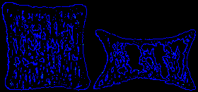
Normal Bone vs. Osteoporotic Bone

How it Happens. Bones are living tissue. Throughout our lifespan, new bone is formed daily to replace areas of bone that dissolve into the blood. This constant remodeling process-bone resorption and then formation-continues throughout life, but after age 35 increasingly less regeneration and more resorption take place. Osteoporosis results when there is excess bone loss without adequate replacement. Bones become brittle and ever more likely to break.
Normal bone structure has two forms. The outer shell of the bone is known as the cortex and is very strong and solid. The inside consists of trabeculae a meshwork of bony struts. The empty spaces between the struts are filled with fat, bone marrow and blood vessels.
In osteoporotic bones, calcium leaches from the bone mass and as a result small holes form in the bones. Presence of these holes causes bone weakening. As the process continues, trabecular struts are lost and the pores and empty spaces within the bone grow larger.
The Shrinking Woman

Initially, only minute breaks may occur in the weakened bone tissue. Eventually, though, major fractures result. As the disease progresses, other characteristics show up: compression of the vertebrae resulting in loss of height and the hunched back deformity known as dowager's hump.
Diagnosis
Back to the Table Of Contents
At present there are as yet no accurate ways to measure bone strength. Bone mineral density measurements are a yardstick for approximately 70% of bone loss. The World Health Organization (WHO) operationally defines osteoporosis as bone density 2.5 standard deviations (SD's) below the mean for young white adult women. (This does not apply to men or children.). (See Figure 4) For advanced osteoporosis, simple X-ray measurements e.g. loss of vertebral height, confirms the diagnosis.
Testing for Osteoporosis
Back to the Table Of Contents
Bone strength is proportional to the amount of bone mineral. Routine x-rays are insensitive to subtle bone loss (it takes a loss of over 30% to show on an x-ray film). Bone mineral can now be measured accurately by a technique called absorptiometry. Radiation absorption is measured to estimate bone mineral. Bone Densiometry has revolutionized the diagnosis of osteoporosis by Dual Photon X-ray Absorption, (e.g. DXA). Radiation exposure is less than with any other medical test involving radiation exposure (less than 1/10th the exposure in a single chest x-ray). A different method of measuring bone mineral uses x-ray and computerized tomography with 25 to 100 times as much radiation per measurement as does gamma ray absorptiometry. It is useful principally for more specialized research measurements of spine mineral response to therapy. Gamma ray absorptiometry may be used to measure bone mineral in the wrist, the hip, the lumbar spine and other sites. Wrist measurements are useful in screening for osteoporosis when combined with spine measurements. Spine measurements are most useful in following bone mineral change due to aging, disease or in response to treatment. Spine mineral tends to show more rapid change than other sites. Hip measurements often show osteoporosis in the hip when other sites are normal. These measurements have led to recognition of so called fracture thresholds below which patients become fracture prone.
Who should have bone mineral measurements?
- Ideally, all white women, and probably all Asian women as well, should be measured at age 45 and then again within a year or two after their menstrual periods have ceased. For moderately severe osteoporosis, the degree of bone loss can be measured and the choice of treatment and response can be followed by serial measurements. This double measurement allows detection of both those with low bone mineral mass at maturity and others who have abnormal rates of bone loss.
- Second, all individuals with multiple risk factors enumerated previously should be measured, regardless of age.
- Third, patients with various diseases and/or who take various medications, e.g. corticosteroids, that may cause osteoporosis should be measured.
| Physiology of Bone Loss | |
|
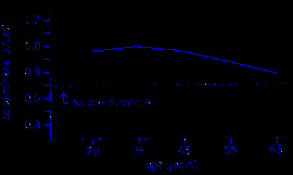
Bone Mineral Density of the Distal Radius (Females) |
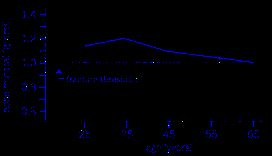
Bone Mineral Density of the Lumbar Spine |
Consequences of Osteoporosis
Back to the Table Of Contents
Some of the many consequences due to osteoporosis are: Increased risk of fracture with minor trauma .. Frequency of traumatic events from lifting and bending impact increases with age, leading to pain and physical incapacity, and loss of income and independence.
The risk of hip fracture for a 50-year-old woman during her lifetime is approximately 14%. Vertebral fractures are more common and very disabling. One-third of patients with hip fractures are discharged to nursing homes and one-fifth live less than one year post fracture (pelvis, ribs etc.). Hip fractures affect quality of life, and some report that 80% of women older than 75 preferred death to a bad hip fracture resulting in permanent nursing home care.
Other quality of life factors include effects on physical health, skeletal deformity, and financial resources. Activities of daily living are affected, as only one-third regain their pre- fracture level of functioning. Depression, fear and anxiety often occur.
Financial Implications:
Direct expenditures for treatment of osteoporosis are 10-15 billion dollars annually, mainly for inpatient care, but do not include treatment for persons without medical insurance. Thus costs are underestimated. Cost of fractures does not include indirect costs, such as loss of wages or productivity. These costs may utilize funds personally allocated for a person's retirement and family needs.
Risk Factors
Back to the Table Of Contents
Primary Osteoporosis
Predictors of low bone mass include being female, increasing age, estrogen deficiency, white race, low weight and body mass index (BMI), family history of osteoporosis, smoking, and prior history of fractures. Consumption of modest alcohol and caffeine-containing beverages are inconsistent risk factors. Level of exercise as children and adolescents are inconsistent and thus are not proven risk factor. Prolonged periods of immobility, early menopause, and low endogenous levels of estrogen appear to play a significant role.
Secondary Osteoporosis
Many disorders are associated with increased risk of osteoporosis: it occurs in 30-60% of cases, such as hypogonadism, (lack of testosterone or estrogens by the testes or ovaries), endocrine disorders, genetic disorders, hematologic disorders, gastrointestinal diseases (such as celiac disease), connective tissue disorders, nutritional deficiency, alcoholism, end stage renal disease, and congestive heart failure. Drug use, such as corticosteroids can have a profound affect. In one study, 10 mg/d of prednisone for 20 weeks resulted in an 8% loss of BMD in the spine. Even inhaled or locally applied corticosteroids may lead to bone loss.
Nutrition
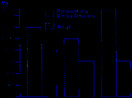
Adequate calcium intake is most important in achieving optimal bone density. The recommended doses of calcium are 800 mg /d age 3-8 and 1300 mg age 9-17. It has been estimated that only 25% of boys and 10% of girls age 9-17 reach these optimal levels. There is a national need to implement adequate calcium intake throughout life, but especially early in life when bone mass is accumulating, during periods of stress, during pregnancy, and especially during lactation, and in old age when calcium absorption is erratic. Adequate Vitamin D intake, i.e. 600-800 IU/day is most important to assure adequate calcium absorption, especially when exposure to ultraviolet light is inadequate. The dietary calcium intake is often underestimated.
Ideal vs. Real Consumption of Calcium

Age: Teenager Premenopausal After Menopause
|
You are welcome to share this © article with friends, but do not forget to include the author name and web address. Permission needed to use articles on commercial and non commercial websites. Thank you. |

